







Eye examination
Content
Description
The eyes work like a perfect camera. The image that is formed on the retina is transmitted to the brain and creates a visual sensation. Just like the camera of your mobile phone, your eyes also require regular care.
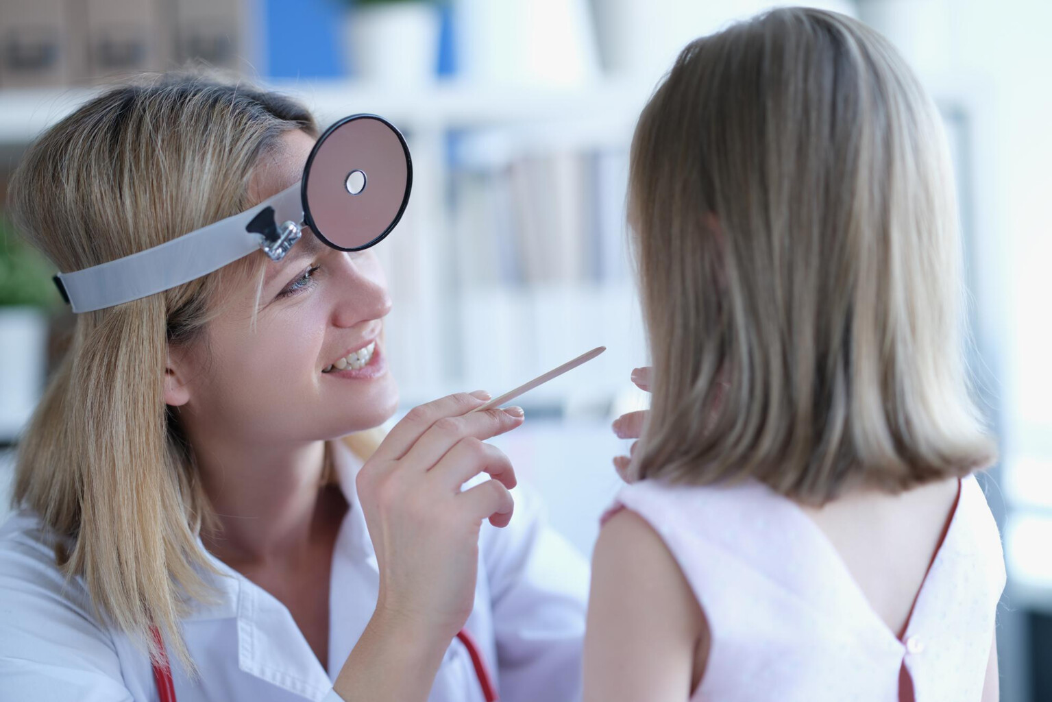
A preventive eye examination should be done every 2 years. It should be remembered that sight is our most important sense. The eye examination takes about 2 hours and the pupil of the eyes must be dilated (using special eye drops). Your vision will therefore be blurred for about 2 - 4 hours.

Facts
- No need for anesthesia
- Outpatient examination
- Examination lasts about 2 hours
- Blurred vision lasts 2 - 4 hours
Basic examinations that reveal the need for glasses and can diagnose eye diseases are:
- Examination of visual acuity (vision) - the patient reads texts of different sizes (near vision) and reads from a blackboard (a Snellen chart) 5 meters away (distance vision). Visual acuity is also measured with a device (autorefractometer).
- Automatic refraction - evaluates refraction, i.e. diopters, which are necessary for reading, foor looking at distance and at medium distance.
- Measurement of intraocular pressure (tonometry) - will measure intraocular pressure, which is important for the diagnos of glaucoma. The device blows a small puff of air on the cornea. At the same time, the thickness of the cornea is measured (pachymetry).
- Slit-lamp - examines the anterior segment (front part) of the eye. That is, the cornea, anterior chamber, iris and lens.
- Ophthalmoscopy - examination of the cornea, lens, retina, macula and optic nerve, vitreous and vessels in the eyes. During this examination the eyedrops are used to dilate pupils.
After the basic examination, the condition of the eyes is evaluated and an overall picture of vision is obtained. If necessary, the eye can be further examined with additional examinations:
- Field of vision (Perimetry) - measurement of the entire field of vision. Each eye is examined separately and visual field gaps are measured.
- Color vision test - a series of images with colored dots is showed to check for color blindness.
- OCT (optical coherence tomography) and fundus photography - examinations (imaging tests) of the individual layers of the retina and optic nerve, which are important in diabetes, glaucoma and age-related macular degeneration.
- Dry eye syndrome - With a lack of tears, the eye does not have sufficient protection (tear film). It is investigated with the Schirmer test and apparatus (IDRA).
- Corneal topography - 3D visualization of the cornea (Pentacam device).
- Endothelial microscope - visualization of the inner wall of the cornea (corneal endothelium).
The ophthalmologist (eye-doctor) may also recommend other examinations or imaging methods - for example USG (ultrasound), Magnetic Resonance Imaging (MRI) and the like.